


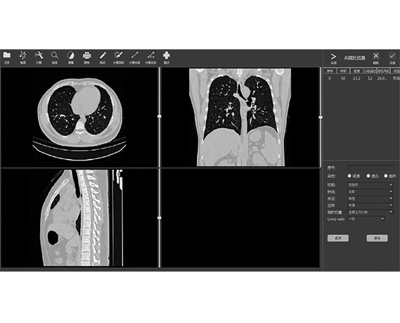
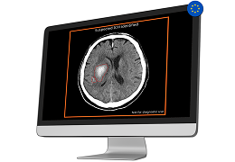
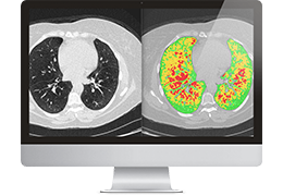
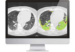
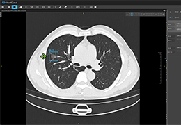
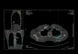
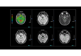
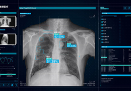
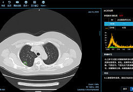
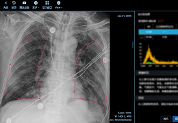
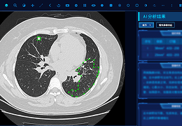
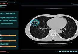
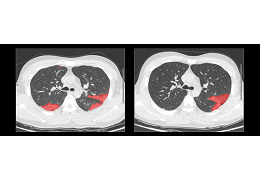
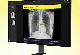
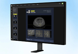
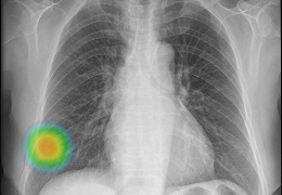
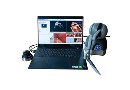
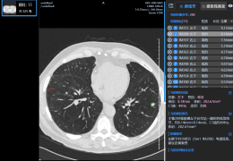
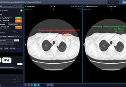
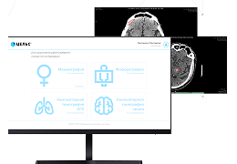
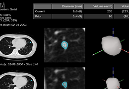
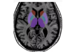
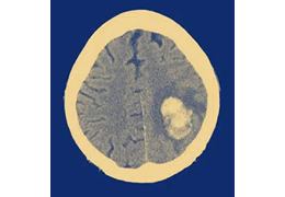
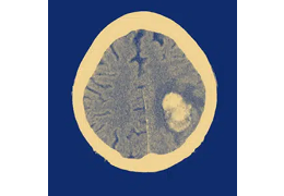
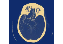
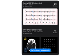
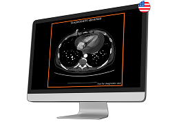
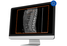
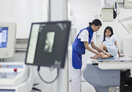
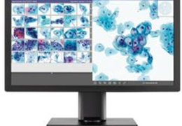
 肺结节智能检测系统
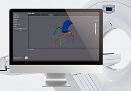
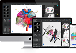
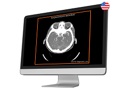
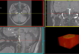
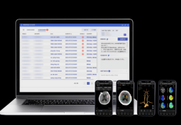
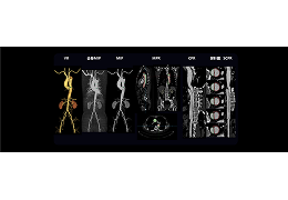
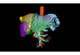
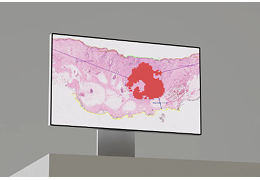
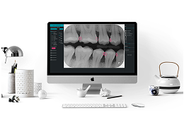
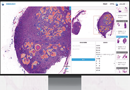
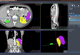
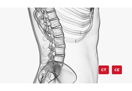
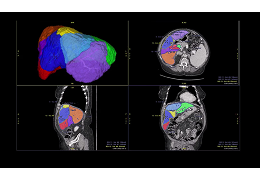
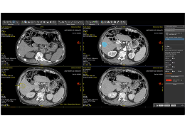
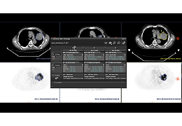
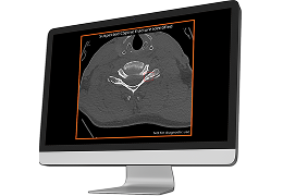
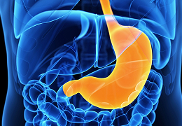
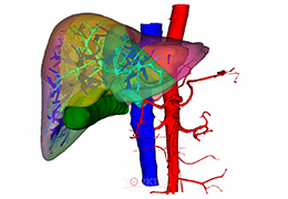 AI+Cloud+3D+ 肝胆系统三维可视化服务
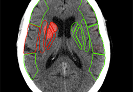
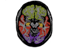
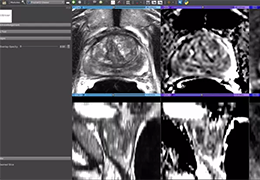
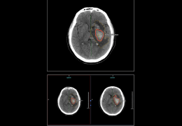
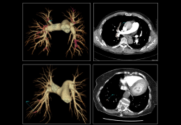
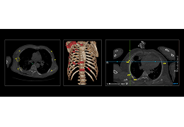
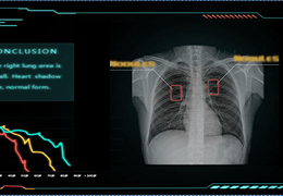
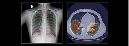
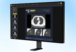
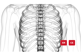
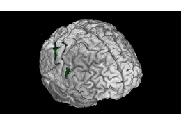
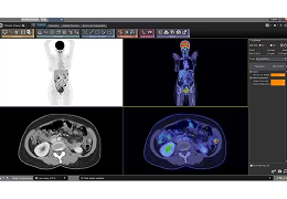
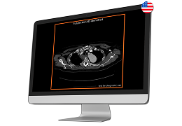
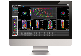
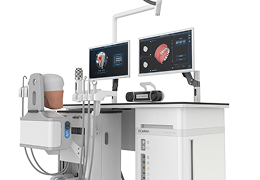
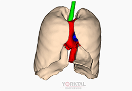 胸肺系统三维可视化服务
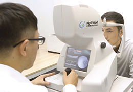
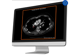
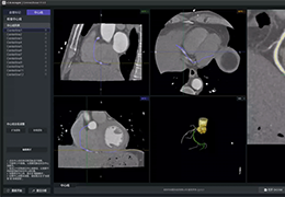
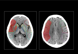
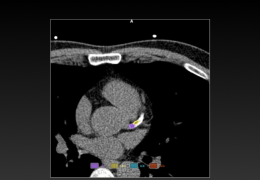
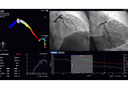
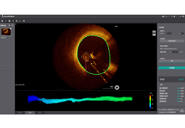
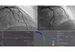
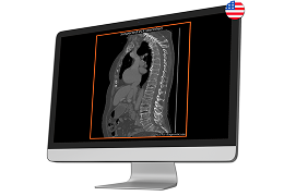
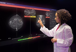
 AccuFFR®ct-基于冠脉CTA的无创FFR分析系统
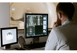
 Al - assisted Diagnosis
 肺癌云网智能筛查平台
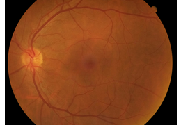
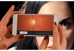
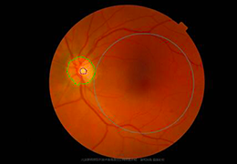 眼底病云网智能筛查平台
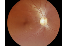
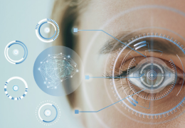
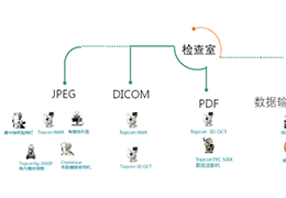 智能眼科影像管理平台
 PV‐iRT 智能放疗辅助系统
 PV-iCS医学影像智能处理系统
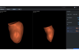
 PV-iSA智能手术辅助系统
 MIAS
MIAS
 睿影AI医学辅助诊断/评估系统
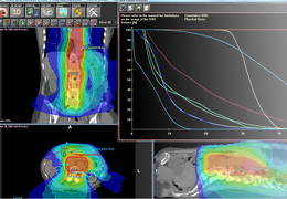
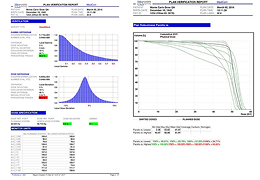
 治疗计划系统RapidPlan
 mdaccAutoPlan®
 影诺智能标注平台
 CINA-ICH
 CINA-LVO
 CINA-ASPECTS
 CINA-PE
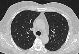 ClearRead-Ct
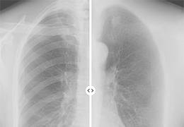 ClearRead Xray
 cmAssist
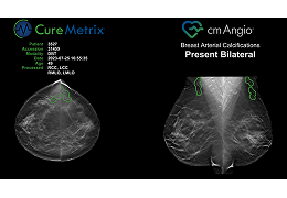 cmAngio
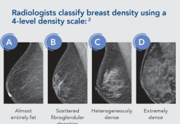 cmDensity
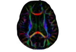
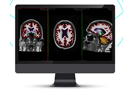
 cNeuro® cMRI
 cNeuro® cDSI
 icobrain ms
 icobrain dm
 icobrain tbi
 icobrain ep
 Lung Density Analysis™ - Inspiration
 Lung Density Analysis™- Functional
 Lung Texture Analysis™
 PANDA
PANDA
 KOALA™
 HIPPO
HIPPO
 LAMA
LAMA
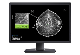
 MammoScreen ®
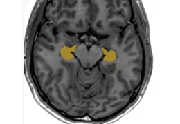
 NeuroQuant
 DeepHealth Prostate
 DeepHealth Brain
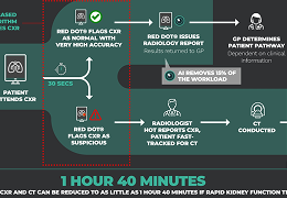
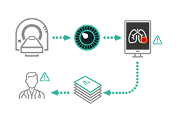 red dot ® Lung Cancer Detection Platform
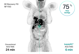 SubtlePET
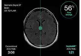 SubtleMR
 MODUS PLAN™
 Celsus. Mammography
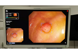
 脉影 CAscope®
 颅影 ICAscope®
 心影 HCscope®
 体素睛采
体素睛采
 体素冠影
体素冠影
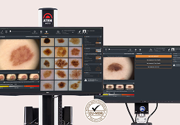
 体素肤知汇
体素肤知汇
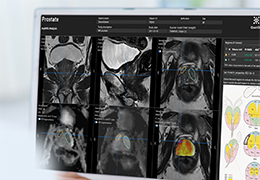
 PROSTATID™
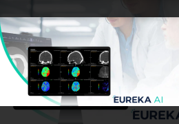 EUREKA
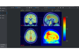
 TR Neuro
 QP-Brain®
 QP-Liver®
 QP-Breast®
 QP-Lung®
 QP-Insights®
 QP-Prostate®
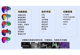
 Al Solutions
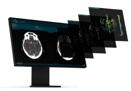 e-CTA
e-CTA
 多参数肝脏健康精准评估
 肝硬化无创评估
 肝肿瘤辅助诊断
 数字医生
数字医生
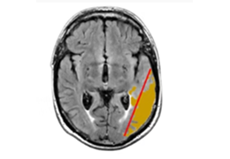
 脑卒中患者出血预测
 脑卒中患者症状智能预测
 帕金森患者自动化分析
 iEcho One-stop
 一栈式智能心脏超声辅助诊断系统
 iNeuroPlan
 致盲眼病AI筛查与评估
 智能影像管理
 近视防控
近视防控
 杏脉锐影 PROXAI
 杏脉镜灵 PYXIS
 放疗信息管理系统RTIS
 放疗勾画工作站
 智能勾画
智能勾画
 CT骨转移智能分析系统
 MR脑转移瘤智能分析系统
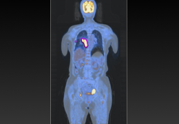 PET/CT智能分析系统
 脑健康数字管理及训练平台
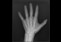
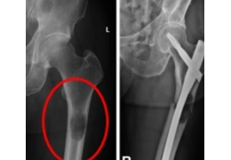
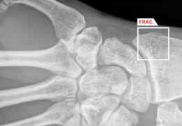
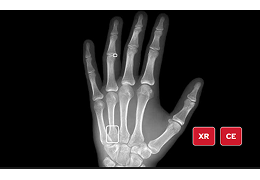
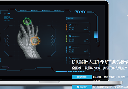 DR骨折人工智能辅助诊断系统
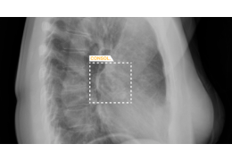
 CT肋骨骨折人工智能辅助筛查系统
 CT病毒性肺炎人工智能辅助筛查系统
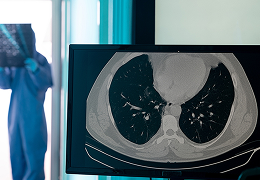
 CT肺结节人工智能辅助筛查系统
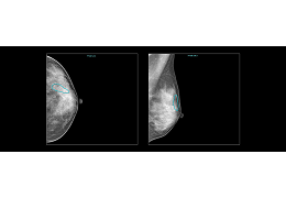
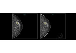
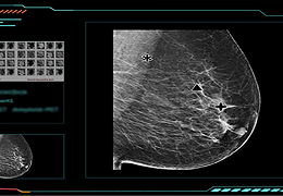
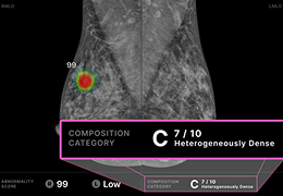
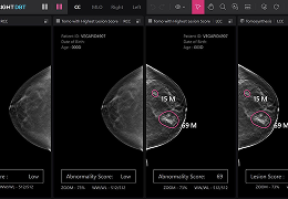
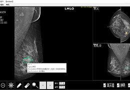
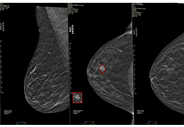
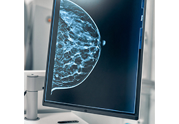
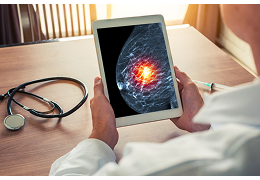
 乳腺钼靶人工智能辅助筛查系统
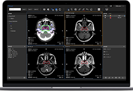
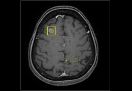
 MR脑结构智能分析系统
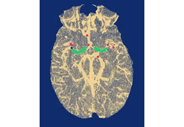
 MR脑小血管病智能分析系统
 CT卒中智能分诊系统
 CT颅内出血智能分析与随访系统
 CT脑缺血智能分析系统
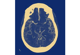
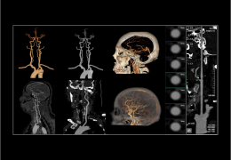 CTA头颈血管智能分析系统
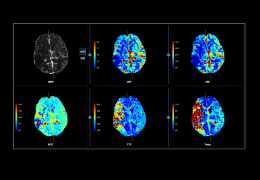 CTP脑灌注智能分析系统
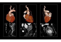 CTA冠脉智能分析系统
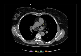 CT 非门控钙化积分智能分析系统
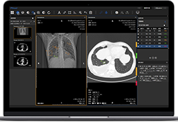
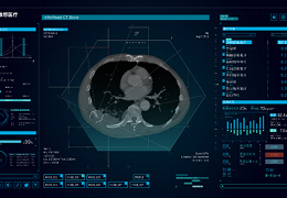
 InferRead CT Lung 肺部疾病智能解决方案
 InferRead CT Fracture 骨疾病智能解决方案
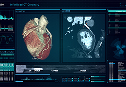 InferRead CTA Coronary 心血管智能解决方案
 InferRead CTA Stroke 头颈CTA智能解决方案
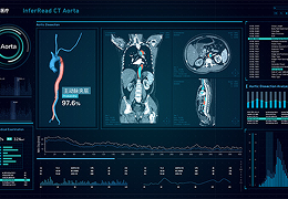 InferRead CTA Aorta 主动脉夹层智能解决方案
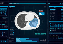 InferRead CT Pneumonia 肺炎疾病智能解决方案
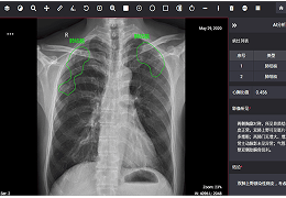
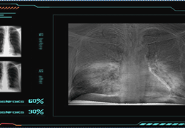
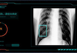
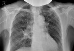
 InferRead DR Chest 胸肺部疾病智能解决方案
 InferRead CT Stroke 脑出血智能解决方案
 CT门控钙化积分智能分析系统
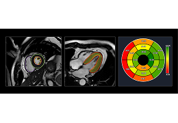 MR心脏智能分析系统
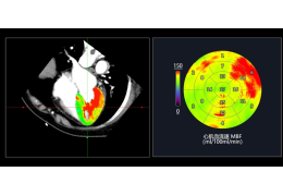 CTP心肌灌注智能分析系统
 CT肺栓塞智能分析系统
 CTA主动脉智能分析系统
 CT骨折智能分析系统
 CT骨转移智能分析系统
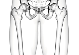
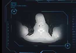
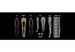
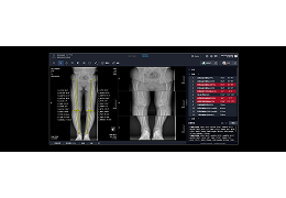 DR下肢力线智能分析系统
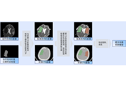
 肝胆手术智能规划系统
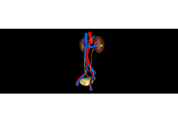 泌尿手术智能规划系统
 CTA下肢血管智能评估系统
 FFDM乳腺智能分析系统
 DBT乳腺智能分析系统
 DR儿童生长发育智能分析系统
 DR肺结核辅助诊断系统
 CT肺结核辅助诊断系统
 结核痰片AI镜检系统
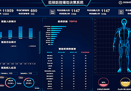 结核防控辅助决策系统
 CT新冠肺炎智能诊断系统
 DR新冠肺炎智能诊断系统
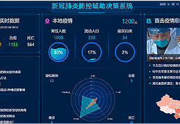 新冠肺炎防控辅助决策系统
 胸部多病种智能诊断系统
 Cancer
 e-ASPECTS
 CT多病种智能诊断系统
 胸片随访对比分析系统
 早期乳腺癌智能诊断及风险评估系统
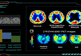 早期老年痴呆症(AD)智能诊断及风险预测系统
 CT早期肺癌结节检测系统
 肺管家胸部健康管理系统
 早期尘肺智能诊断及风险评估系统
 胸部正异常筛查系统
 智能胸部体检诊断系统
 CT肺炎智能分析系统
 影像检查智能质控系统
 肿瘤靶区与危及器官自动勾画系统 DeepContour
 心电脑卒中智能诊断及风险评估系统
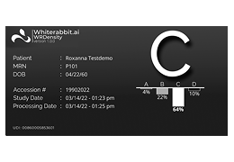 WRDensity
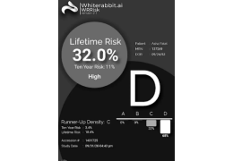 WRRisk
 PROGNICA MMG
 DeePathology®STUDIO
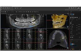
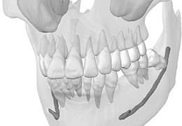
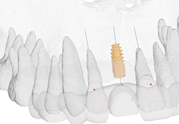
 Simfini-IMPLANT
 Simfini-TUMOR
 Simfini-OSTEO
 AZtrauma
 AZchest
 AZmeasure
 AccuFFRct-基于CTA的无创FFR技术
 AccuFFRangio-基于冠脉造影的FFR技术
 AneuAccuFFRivus-基于IVUS的FFR技术
 AccuFFRoct-基于OCT的FFR技术
 ProSoma
 ProSoma Core
 AccuLV-心脏无创功能学评估产品
 AcculMR-微循环阻力评估系统
 ARPlanner®
 mdaccAutoPlan®
 Cleo Breast
 cleoskin
 RADIOLens
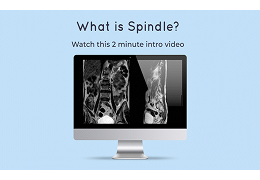 Spindle
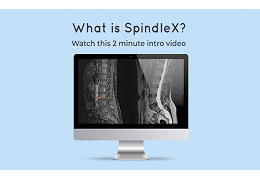 SpindleX
 Teleradiology
 GleamerBoneView
 BoneViewBoneMetrics
 BoneViewBoneAge
 GleamerChestView
 GleamerBreastView
 GleamerLumbarMR
 Pixyl.Neuro
 GleamerBoneCT
 GleamerLungCT
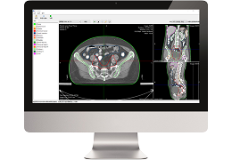
 IMAGING MANAGEMENT SYSTEM
 IMAGING BIOMARKERS
 DeepDx® Prostate – CNB
 DeepDx® Prostate – RP
 DeepDx® Prostate – TURP
 DeepDx® Breast – Resection
 DeepDx® Breast – SLNB
 Lunit INSIGHT CXR
 Lunit INSIGHT MMG
 Lunit INSIGHT DBT
 Lunit SCOPE IO
 Lunit SCOPE PD-L1
 LunIt SCOPE HER2
 Lunit iNSICHT CXRTriage
 Lunit SCOPE GP
 HTG
HTG
 VOXEL-MAN Sonography
 DentaliQ caries
 OptaliQ glauco
 爱昔瞳TM视网膜人工智能筛查
 PRODUCT SERVICES
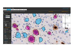
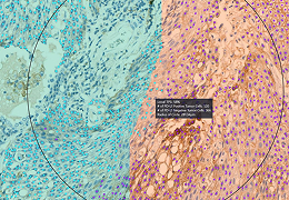
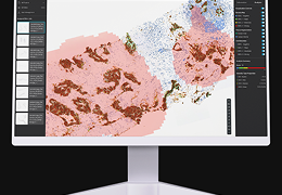
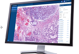
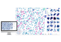
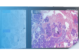
 CellPlatform 数智病理AI辅助诊断系统
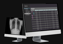
 iRay DR
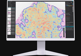
 CT智能辅助诊断系统
 DR常见病智能筛查系统
 VistaSoft 2D Xray
 VistaSoft 3D Xray
 VistaSoft Trace
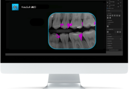 VistaSoft AID
 PaxeraUltimaAi
 Liflow
 Logicon Caries Detector Software Version 5.2
 Advantis Prostate
 Second Opinion® AI
 CHIMAERA AI-B2
 AI – WELL PREPARED
 AIMEE
AIMEE
 Moleanalyzer pro
 handyscope pro App
 CBCTSegmentation
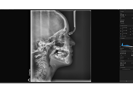
 Brain computed tomography
 Fluorography and chest x-ray
 AI
AI
 Additional software
 red dot® CTH V1
 RadOS
RadOS
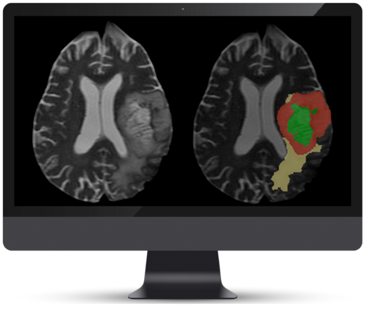
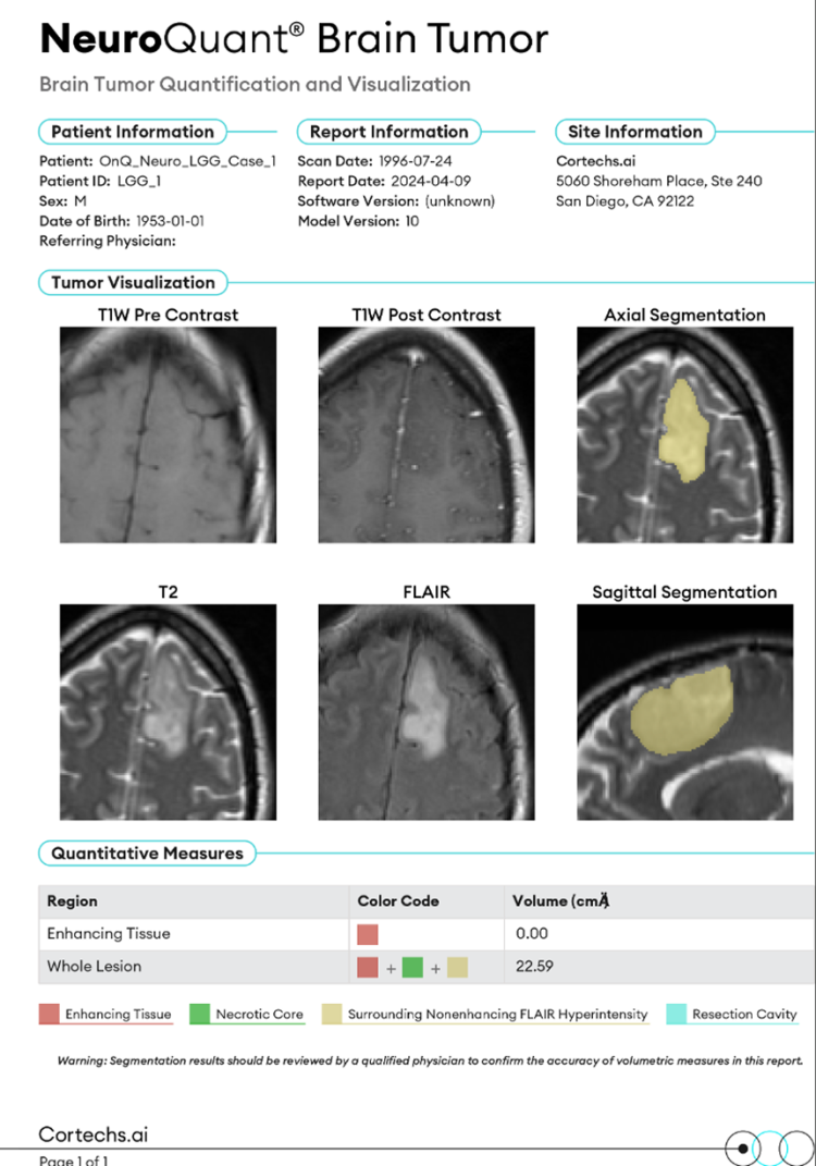
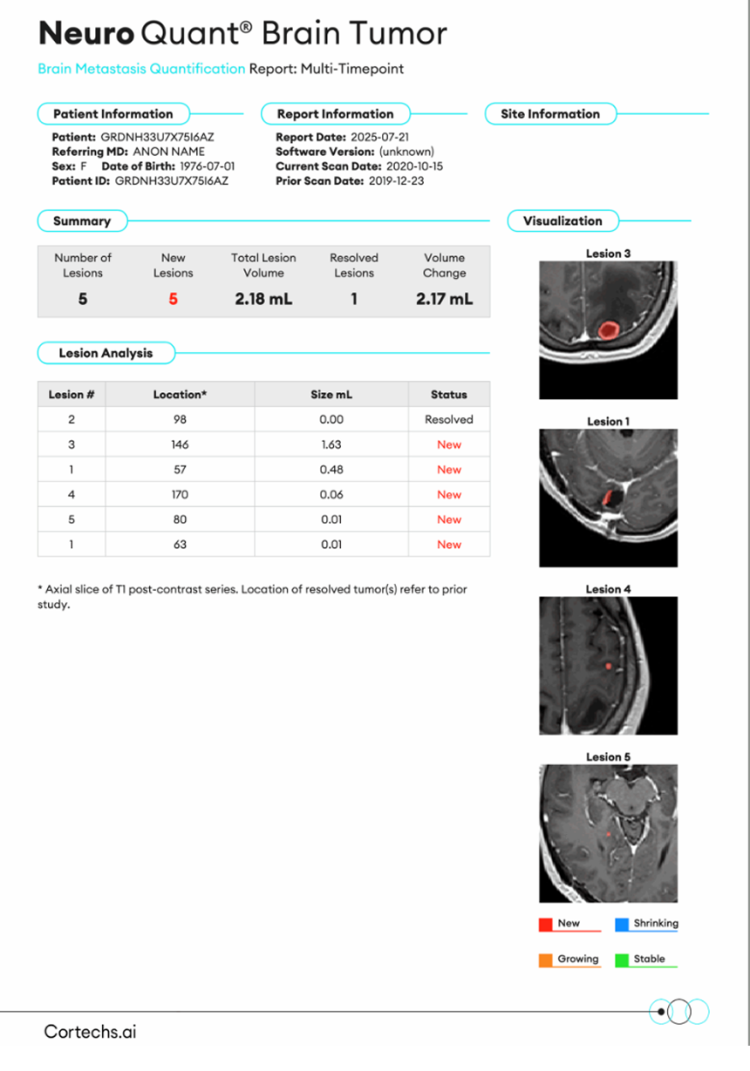
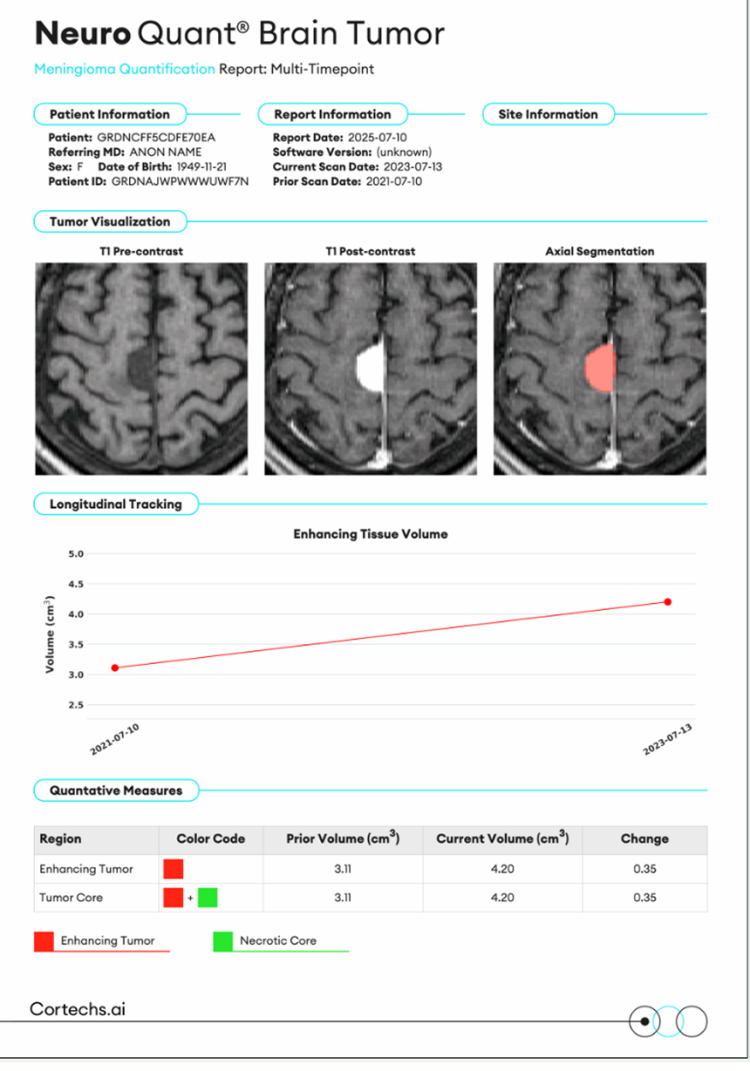
 NeuroQuant Brain Tumor®
 NeuroQuant PET®
 OnQ™ Prostate
 NeuroQuant MS®
 NeuroQuant for ARIA®
 NeuroAlign CT
 DeepHealth Lung
 SmartMammo™
 Koios DS™ Breast
 Koios DS™ Thyroid
 IB Lab SQUIRREL™
 IB Lab FLAMINGO
 RBfracture™
 icobrain aria
 icobrain pd
 BrainWave
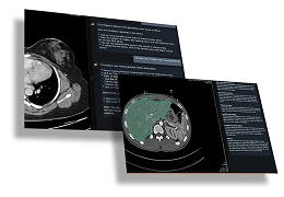
 Hepatic VCAR
 Liver Suite
 OncoQuant
 PET VCAR
 Viz Aneurysm
 Viz CTP
 Viz Hemorrhage
 Viz ICH+
 Viz LVO
 Viz Connect®
 Viz Aortic
 Viz PE
 Viz HCM
 cNeuro® cPET and cDAT
 CINA-iPE
 CINA-AD
 CINA-CSpine
 CINA-VCF
 CINA-VCF Quantix
 乳腺智能检测云
 临床决策支持系统CDSS
 Concentrig AP
 Concentrig AP-DX
 Concentrig LS
 Automated QC
 Real-World Data
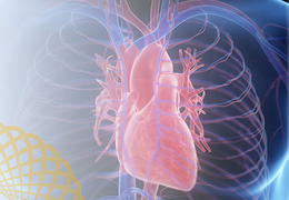 AI Cardiac Solution
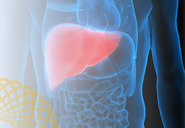 AI Liver Solution
 Nanox.AI
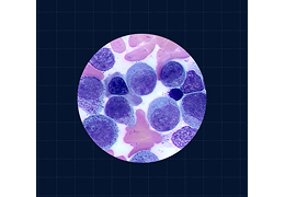 Full-Field Bone Marrow AspirateTM (FF-BMA) Application
 Paige Prostate
 Paige GI
 Paige PanCancer
 肺结核X射线图像辅助评估软件
 CBCT Segmentation
 Implant Guide Design
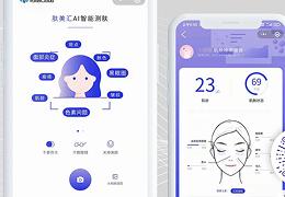 体素肤美汇
体素肤美汇
 天眼智能肿瘤靶区勾画系统
 天玑智能放疗工作站
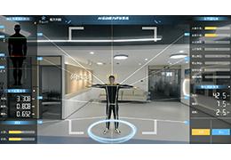 康复智能评估系统
 影诺鹰眼辅助诊疗系统
 胸部CT肺炎智能分析系统
 肺部智能手术规划系统
 肺部智能穿刺消融规划系统
 肺部智能气管镜规划系统
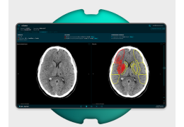
 深脉灵析:头颈智能辅助诊断系统
 头颈血管智能分析系统
 脑出血智能分析系统
 脑缺血智能评分系统
 脑灌注智能分析系统
 肝脏MR智能分析系统
 肝脏智能手术规划系统
 肝脏智能穿刺消融规划系统
 骨龄智能分析系统
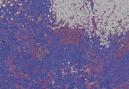
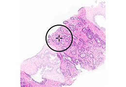
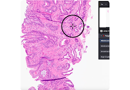
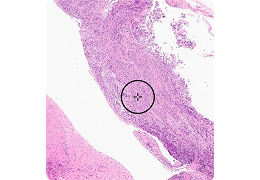
 消化道病理智能分析系统
 宫颈TCT智能分析系统
 乳腺免疫组化智能分析系统
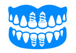
 艾知星 口腔技能训练及实时评估系统
 Genius AI® Detection PRO Assistant
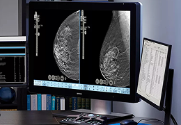 Hologic Genius AI® Detection Solution
 肺部健康精准评估报告系统
 ZBEdge® Dynamic Intelligence™
 Genius™ Cervical AI
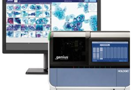 Genius™ Digital Diagnostics System
 Genius™ Review Station
 心电血压数据管理系统
 人工智能辅助宫颈癌诊断系统
 人工智能辅助胃癌诊断系统
 人工智能辅助甲状腺癌诊断系统
 KFBIO 远程病理学诊断平台
 ProFound Detection V4
 ProFound AI® V3
 SecondLook®
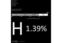 ProFound AI Risk
 NDI Software Packages