FastStroke
Bringing automation to your CT Stroke workflow and assisting with communication between care team members.
At a glance
0-click workflow
Background processing of CT ischemic stroke work up
Automated email
Automatically send preprocessed images and functional maps to stroke team¹
AI‑based processing
Automated Large Vessel Occlusion (LVO) detection,
powered by StrokeSENS²
Automate the work up of CT ischemic stroke cases
A comprehensive workflow solution, FastStroke can automatically process and send images to the stroke team via email¹. It also offers an option to seamlessly integrate with Circle Neurovascular’s StrokeSENS™ AI‑based processing tool for LVO detection².

0-click workflow
Background processing of LVO detection², mCTA and Perfusion³
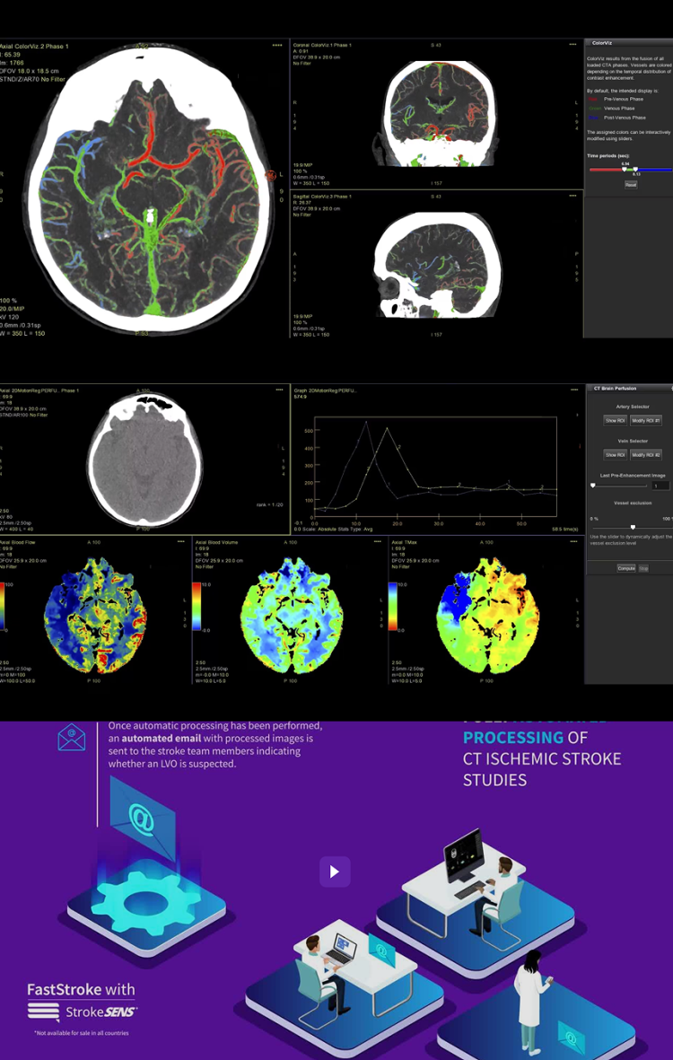
Auto sends preprocessed images, LVO finding, functional perfusion maps and results in email¹ format to stroke team
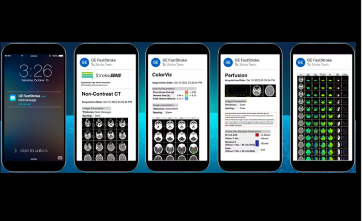
Seamless integration to Circle Neurovascular's StrokeSENS² AI-based processing tools
StrokeSENS LVO²
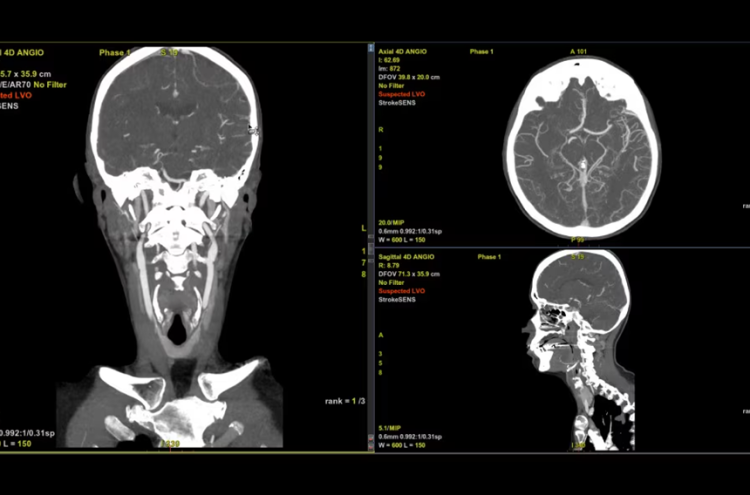
StrokeSENS LVO uses AI to identify Large Vessel Occlusion (LVO) on CT Angiography.
StrokeSENS LVO automatically notifies the stroke team of suspected LVO cases within minutes of receiving the CTA image, supporting early engagement of the stroke team.
In the case a LVO is detected, Suspected LVO is written onto the input DICOM image (CTA or 1st phase of mCTA) and results are automatically included in the email notification, helping notify the stroke team of the time-sensitive case.
Interactive workflow
Intuitive workflow to quickly move through all acquired series
Intelligent loading identifies the series type and applies the appropriate layout and protocol
Automatic synchronization of phases from multiphase CTA (mCTA)
Smart layouts automatically adjust to display up to 6 mCTA phases simultaneously
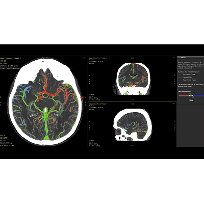
CTA images are automatically displayed in a thick 2D MIP at optimized WW/WL settings
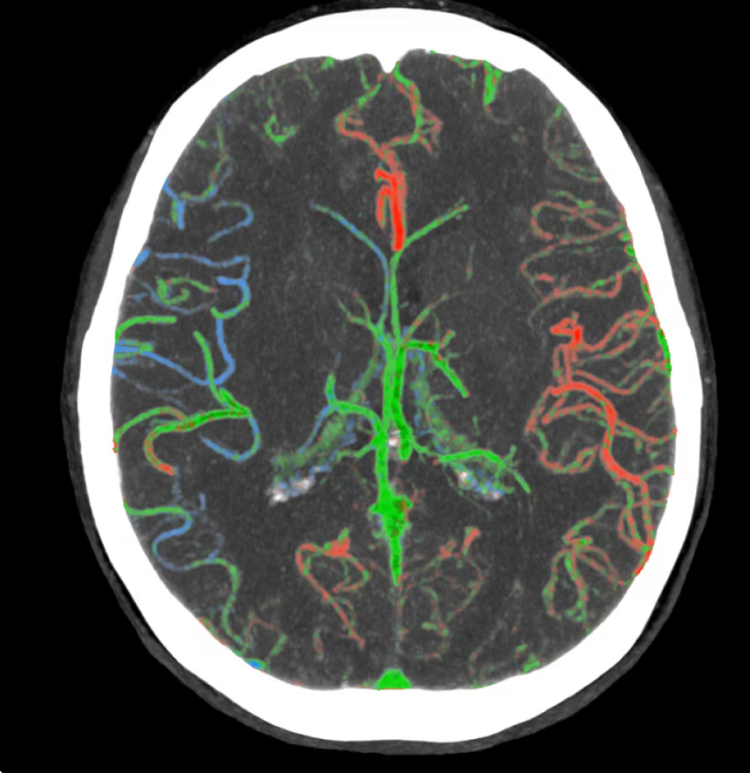
ColorViz, an intelligent color-coded display enabling easy and confident identification of vascular enhancement timing
Perfusion maps and tissue classification
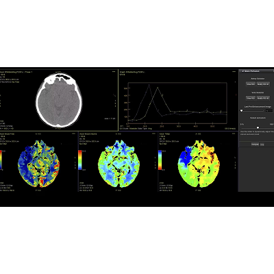
Fully integrated with CT Perfusion 4D for visualization of perfusion functional maps³
Deep Learning brain ventricle segmentation to prevent ventricular matter inclusion in quantitative results and improve visual inspection of the maps
Automated computation of the functional maps
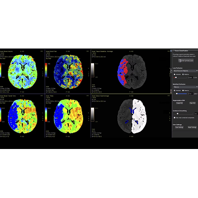
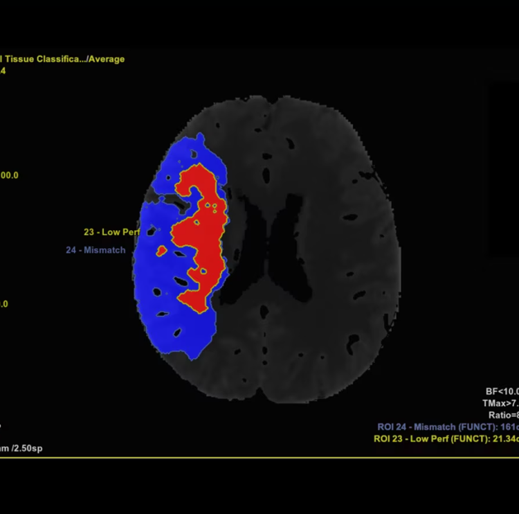
Tissue Classification map segmented from absolute or relative values, customizable thresholds and user selectable input maps
Mismatch volume and ratio calculated from the Modified Perfusion region and the Low Perfusion region
Related documents
Clinical white paper: Tissue Segmentation in Acute Ischemic Stroke
References
1. Available on AW Server. The email license may not be available in all countries or regions. Warning: This email is not intended for primary diagnosis. See PACS or dedicated review station for diagnostic interpretation of results. Warning: This email was generated automatically without prior user review.
2. StrokeSENS™ is legally manufactured by Circle Neurovascular Imaging, Inc. StrokeSENS license is a pre-requisite for StrokeSENS’ LVO within FastStroke. Not available for sale in all countries.
3. CT Perfusion 4D License is a pre-requisite for Neuro Perfusion maps calculation within FastStroke.

非常抱歉,您只有购买软件后才能查看完整软件教程!
